MDCAT Biology – Chap#5 Coordination and Control
Nervous system
The body of an animal is frequently exposed to variety of stimuli in its daily life. For an appropriate response to a particular stimulus, usually more than one body parts are involved, their activities are coordinated either by nervous system or endocrine system or both.
Nervous Coordination
This type of co-ordination involves specialized cells or neurons linked together directly or indirectly via the central nervous system, to form network that connects the cell or organs which receive stimuli and those which carry out actions or responses.
The neurons have the capacity to generate and conduct impulses which travel across the synapse.
Three basic components of nervous system are:
- Receptors
- Neurons
- Effectors
Sensory Receptors and Their Working
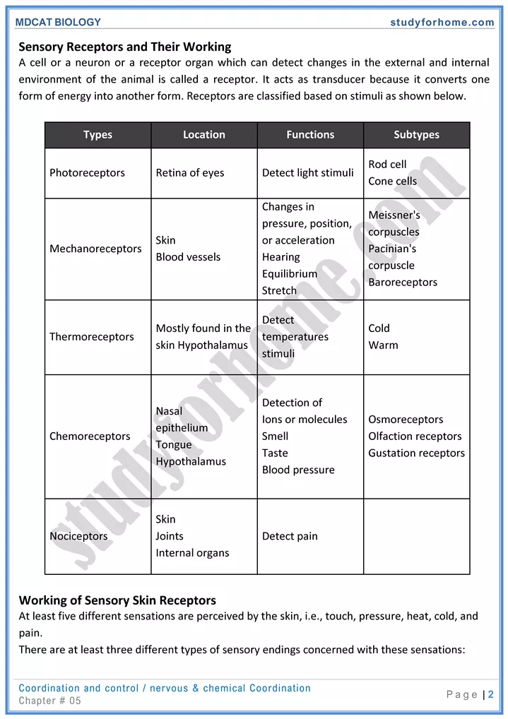
A cell or a neuron or a receptor organ which can detect changes in the external and internal environment of the animal is called a receptor. It acts as transducer because it converts one form of energy into another form. Receptors are classified based on stimuli as shown below.
| Types | Location | Functions | Subtypes |
| Photoreceptors | Retina of eyes | Detect light stimuli | Rod cell Cone cells |
| Mechanoreceptors | Skin Blood vessels | Changes in pressure, position, or acceleration Hearing Equilibrium Stretch | Meissner’s corpuscles Pacinian’s corpuscle Baroreceptors |
| Thermoreceptors | Mostly found in the skin Hypothalamus | Detect temperatures stimuli | Cold Warm |
| Chemoreceptors | Nasal epithelium Tongue Hypothalamus | Detection of Ions or molecules Smell Taste Blood pressure | Osmoreceptors Olfaction receptors Gustation receptors |
| Nociceptors | Skin Joints Internal organs | Detect pain |
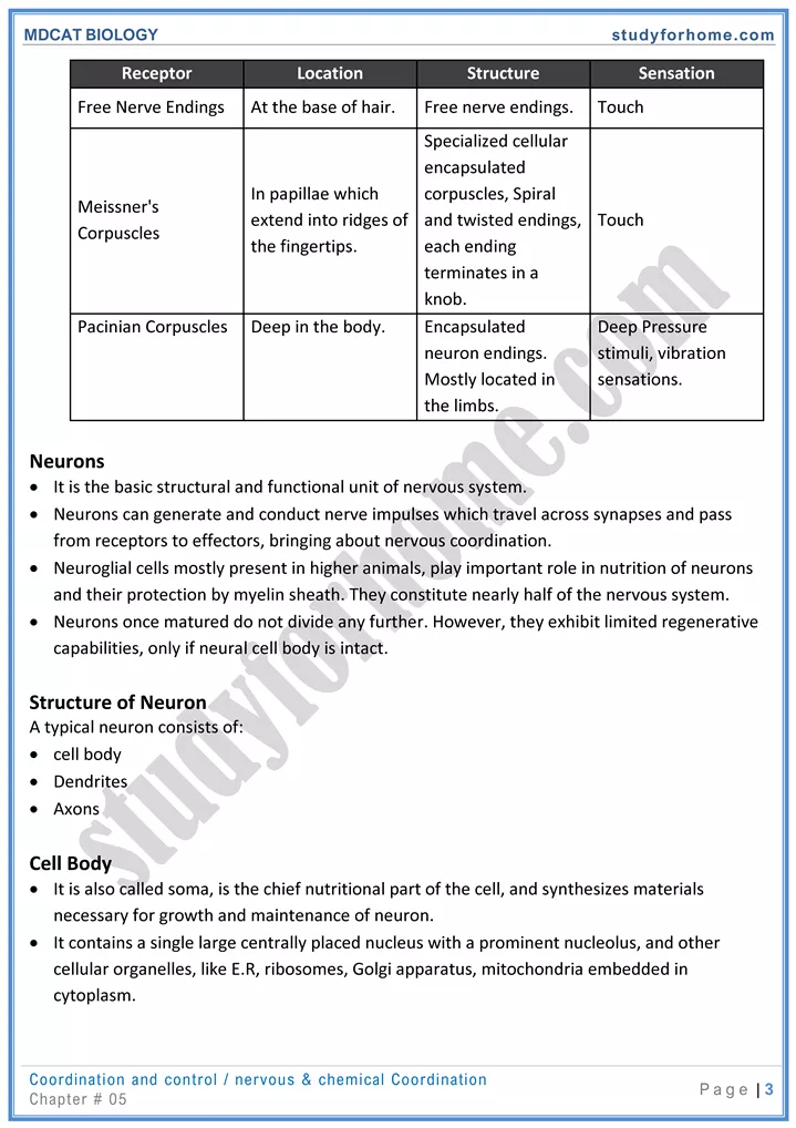
Working of Sensory Skin Receptors
At least five different sensations are perceived by the skin, i.e., touch, pressure, heat, cold, and pain.
There are at least three different types of sensory endings concerned with these sensations:
| Receptor | Location | Structure | Sensation |
| Free Nerve Endings | At the base of hair. | Free nerve endings. | Touch |
| Meissner’s Corpuscles | In papillae which extend into ridges of the fingertips. | Specialized cellular encapsulated corpuscles, Spiral and twisted endings, each ending terminates in a knob. | Touch |
| Pacinian Corpuscles | Deep in the body. | Encapsulated neuron endings. Mostly located in the limbs. | Deep Pressure stimuli, vibration sensations. |
Neurons
- It is the basic structural and functional unit of nervous system.
- Neurons can generate and conduct nerve impulses which travel across synapses and pass from receptors to effectors, bringing about nervous coordination.
- Neuroglial cells mostly present in higher animals, play important role in nutrition of neurons and their protection by myelin sheath. They constitute nearly half of the nervous system.
- Neurons once matured do not divide any further. However, they exhibit limited regenerative apabilities, only if neural cell body is intact.
Structure of Neuron
A typical neuron consists of:
- cell body
- Dendrites
- Axons
Cell Body
- It is also called soma, is the chief nutritional part of the cell, and synthesizes materials necessary for growth and maintenance of neuron.
- It contains a single large centrally placed nucleus with a prominent nucleolus, and other cellular organelles, like E.R, ribosomes, Golgi apparatus, mitochondria embedded in cytoplasm.
- The cytoplasm is characterized by the presence of Nissl’s granules which are group of ribosomes associated with RER and Golgi apparatus.
- If it is intact, the neuron can regenerate its axonal and dendrite components.
Dendrites
- These are processes that carry impulses towards the cell body.
- These are usually short, thin, often highly branched cytoplasmic extensions and devoid of Schwann cells and thus non-myelinated.
Axons
- Axons of other neurons form synapses with the dendrites. They give a spiny look to the neurons.
- The processes carrying impulses/action potentials away from cell body are called axons.
- An axon is comparatively a long and thick nerve fibre which has a constant diameter and can vary in size from a few millimeters to a meter length.
- It may be branched or unbranched. It terminates by branching to form small extensions with enlarged ends, called pre-synaptic terminals.
- Cellular organelles like mitochondria, microtubules and neurofibrils, R.E.R. and GA are present throughout the axoplasm of the neuron.
- Most of the axons are surrounded by protective sheaths called myelin sheath, which is important for neuronal nutrition, protection and proper propagation of impulses.
Types of Neurons
All neurons vary somewhat in size, shape, and characteristics depending on the function and the role of the neuron. Based upon function, there are three types of neurons:
- Sensory Neurons
- Motor Neurons
- Associative Neurons
Sensory Neurons
Sensory neurons carry sensory information from receptors to associative neurons present in CNS.
The dendrite endings of some sensory neurons also act as receptors.
They usually have single long dendrite called dendron. It is structurally and functionally similar to axon.
Motor Neurons
Motor neurons conduct impulses away from the central nervous system. The dendrites make contact with other neurons in the spinal cord. The terminal branches at the far end of the neuron are connected to an effector.
Associative Neurons
Associative (intermediate/relay/inter/connector) neurons occur entirely within the CNS and connect sensory and motor neurons.
The axon is comparatively thin and non-myelinated.
They are involved in processing and interpretation of information coming from receptors.
Nerve impulse
Nerve impulse is a wave of electrochemical changes, which travels along the length of neurons involving movement of ions across the membrane and chemical reactions.
Initiation of Nerve Impulse
- A stimulus capable to produce action potential in neurons is call threshold stimulus and if it is not capable to excite neurons, then it is called sub-threshold stimulus.
- It results in a remarkable localized change in the resting membrane potential. It disappears for a brief instant and is replaced by action potential. This change is so brief (for a millisecond) that only a portion of neuron is in active state.
Repolarization of Neuron (AMP —i RMP)
- It is the restoration of resting membrane potential, after the wave of depolarization has passed.
- It results from closure of Na+ gates and opening of K+ gates, therefore K+ diffuses out of the cell. Since K+ is positively charged, this makes the inside of the cell more negative and starts the process of repolarization.
Generation and Transmission of Nerve Impulse
Electrochemical word refers to the electrical potential that exists on neuron membrane. In case of neuron, the electrical potential is termed as membrane potential which is exhibited in two different forms i.e., resting membrane potential (RMP) and active membrane potential (AMP).
Resting Membrane Potential
- Potential difference across the membrane when neuron is in not being stimulated/non-conducting state is called resting membrane potential, and is characterized by more positive outside surface of neuron membrane than inner surface.
- Neuron in this state is in polarized form.
Active Membrane Potential/ Action Potential
- AMP is characterized by more positive inside of neuron than outside. This happens when positive charges tend to move inside of neuron on receiving a particular stimulus.
- This electrochemical change appears on a short region of neuron for a brief period of time followed by the recovery of the polarized state.
Synapse:
The junction between axon terminal of one neuron and the dendrite of another neuron, where information from one neuron is transmitted or relayed to another neuron is called synapse.
Structure of Synapse
The neurons are not in direct contact at a synapse. There is a gap, called a synaptic cleft between them.
A single neuron may form synapses with many incoming fibres of different neurons.
A neuron which carries an impulse toward a synapse is called pre-synaptic neuron while the one which receives the impulse after it crosses the synapse is a post synaptic neuron.
A single nerve impulse does not necessarily get across the synapse. It may take two or three impulses arriving in rapid succession or perhaps simultaneously from two or more fibers to start an impulse in the next neuron.
Mechanism of Synaptic Transmission
Synaptic transmission takes place in the form of a message which is transmitted across the synapse in the form of chemical messengers called neurotransmitters.
The axons usually have several rounded synaptic knobs which contain numerous membranous sacs, called synaptic vesicles.
The main steps involved in synaptic transmission is shown in the following diagram:
There are two types of synapses;
- Electrical synapses
- Chemical synapses
Electrical synapses
In electrical impulse, which are specialized for rapid signal transmission, the cells are separated by a gap, the synaptic cleft, of only 0.2nm, so that an action potential arriving at the pre synaptic side of cleft, can sufficiently depolarize the post synaptic membrane to trigger its action potential directly.
Chemical synapses
The majority of synapses are chemical synapse where synaptic cleft has gap of more than 20nm.
Through these synapses, information of impulse from one neuron is transmitted to another by means of chemical messengers, the neurotransmitters.
Spinal Cord
The spinal cord is the most important structure between the body and the brain.
The spinal cord extends from the foramen magnum (a hole in the bottom of skull) where it is continuous with the medulla to the level of the first or second lumbar vertebrae.
It is a vital link between the brain and the body, and from the body to the brain.
Structure
A transverse section of the adult spinal cord shows white matter in the periphery, grey matter inside, and a tiny central canal filled with CSF at its center.
Grey Matter
Grey matter is shaped like the letter ‘H or a ‘butterfly’.
As in the other part of the nervous system, the grey matter consists of neuron cell bodies and non-myelinated parts of the fibres.
Several pairs of spinal nerves originate from ventral horn of grey matter. Similarly, several pairs of spinal nerves join the spinal cord through dorsal horn of grey matter.
Dorsal root of spinal nerves, also contain ganglia present just beside the spinal cord.
White Matter
The white matter is made up of bundles of myelinated fibres.
White matter shows deep grooves from both side i.e., from dorsal side to the central canal and from ventral side to the central canal.
Segments of Spinal cord
The spinal cord is divided into four different regions: the cervical, thoracic, lumbar and sacral regions.
The different cord regions can be visually distinguished from one another. Two enlargements of the spinal cord can be visualized: The cervical enlargement, which extends between C3 to T 1; and the lumbar enlargements which extends between L1 to S2
The cord is segmentally organized. There are 31 segments, defined by 31 pairs of nerves exiting from the cord. These nerves are divided into 8 cervical, 12 thoracic, 5 lumbar, 5 sacral, and I coccygeal nerve. Dorsal and ventral roots enter and leave the vertebral column respectively through intervertebral foramen at the vertebral segments corresponding to the spinal segment.
Online MCQs Test of Coordination and Control
Preparation of Multiple Choice Questions of Coordination and Control