Life process in Animals and Plants – Chap 9 – Biology MDCAT
Nutrition
Carnivorous Plants
-
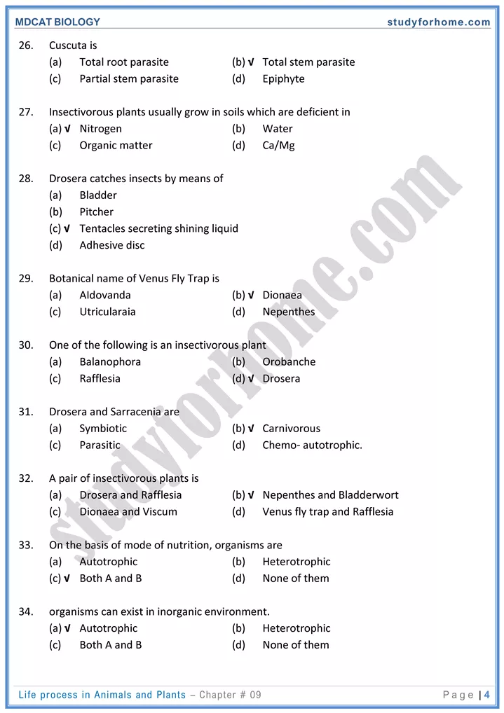
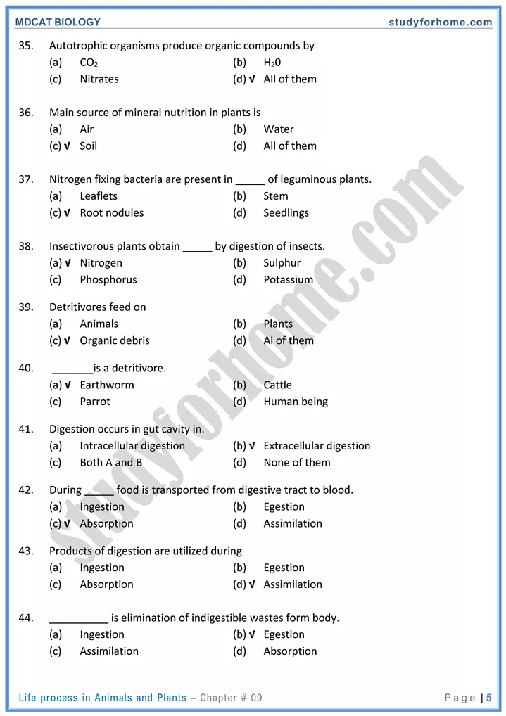
- There are a few plants that supplement their inorganic diet with organic compounds, These organic compounds are obtained by trapping and digesting insects and small animals.
- All of the insectivorous plants are true autotrophs, but when they capture prey, their growth becomes rapid.
- Apparently, nitrogenous compounds of animal body are of benefit to these plants. In some plants, the trapped insects are decomposed by bacteria.
- In others, the trapped insects are digested by enzymes secreted by the leaves. The plants absorb the nitrogenous compounds thus formed.
Digestive System
-
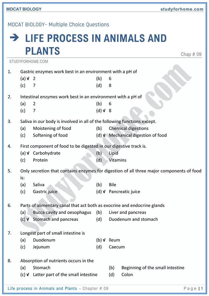
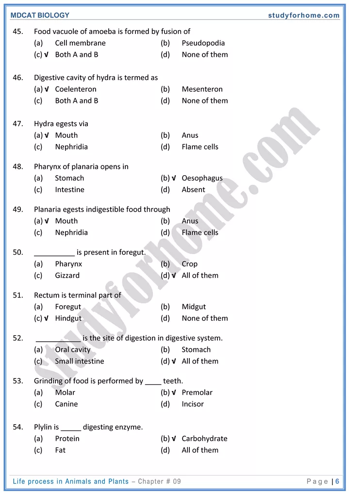
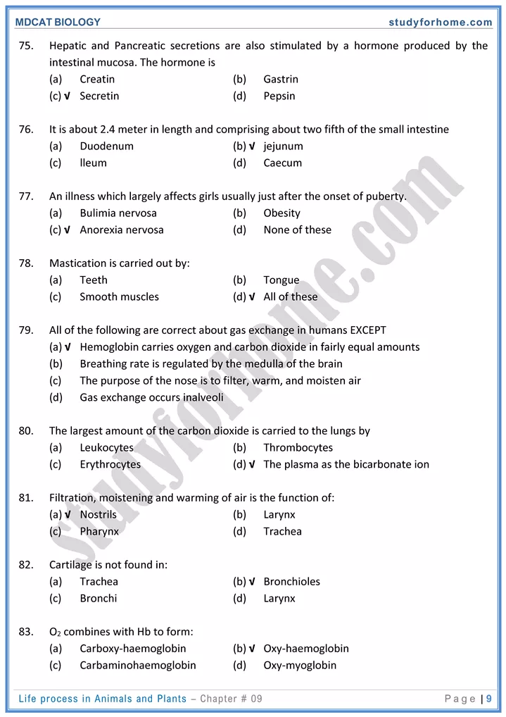
- Anatomically and functionally, the digestive system can be divided into alimentary canal/GIT and accessory digestive structures. The structures of GIT include oral cavity, pharynx, esophagus, stomach, small intestine, and large intestine. The accessory digestive structures include the teeth, tongue, salivary glands, liver, gall bladder and pancreas.
- The GIT consists of a continuous long coiled tube that extends from mouth to anus.
- It is specialized at various points along its length, with each region designed to carry out a different role. The GIT is approximately 9m/30ft long.
- The digestive tube consists of four major layers: an internal mucosa, sub-mucosa, muscularis and an external serosa. These four layers are present in all areas of the digestive tract from esophagus to the anus.
- The main parts in the direction of passage of food are:
- Oral/buccal → cavity Esophagus → Stomach → Small Intestine (Duodenum → Jejunum → Ileum) → Large Intestine (Caecum → Ascending Colon → Transverse Colon → Descending Colon → Sigmoid Colon → Rectum)
- The salivary glands, liver, and pancreas are hot part of the digestive tract, but they have a role in digestive activities and are considered accessory glands.
Digestion in Oral Cavity
-
- The mouth is surrounded by the lips, cheeks, tongue and a palate and includes a chamber between the palate and tongue called oral cavity. The tongue nearly fills the oral cavity when the mouth in closed. It is the site for entrance of food in alimentary canal.
- Rough projections called papillae on the surface of the tongue cause friction; which is useful in handling of food. These papillae also contain taste buds.
- The palate forms the roof of the oral cavity, It consists of a hard anterior part the hand palate and soft posterior part, the soft palate.
Functions of Oral Cavity
It performs four important functions:
-
- Selection of food
- Grinding or mastication of food
- Lubrication of food
- Enzymatic digestion of food
Selection of Food:
-
- When food enters in oral cavity, it is tasted, smelled and felt. If the taste or smell is unpleasant or if hard objects like bone or dirt are present in the food, it is rejected.
- Oral cavity is aided in selection by the senses of smell, taste and sight.
- Tongue being sensory and muscular organ plays the most important role in the selection of food through its taste buds.
Grinding or Mastication:
-
- After selection, the food is ground by means of molar teeth into small pieces. This is
- useful because:
- Esophagus allows relatively small pieces to pass through.
- Small pieces have much more surface for enzymes to attack.
Pharynx:
-
- The pharynx is a cavity behind the mouth. It is common passage for digestive system and
- respiratory system. It is lined by mucus.
Swallowing:
-
- As a result of mastication, softened, partly digested and slimy food mass is rolled into small oval lump called bolus, which is then pushed to the back of the mouth by the action of tongue and muscles of pharynx.
- Transfer of bolus from buccal cavity to pharynx and then to esophagus is called swallowing/ deglutition.
- Beginning of swallowing is voluntary action and then it becomes involuntary. The swallowing procedure is regulated by nerves in the medulla oblongata and pons.
Events of Swallowing:
-
- Tongue moves upwards and backwards against the roof of mouth, forcing the bolus to the back of the mouth cavity.
- Soft palate is pushed up by tongue which closes nasal opening at the back.
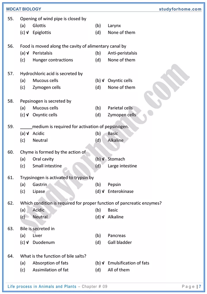
- Tongue forces the epiglottis (flap of cartilage) into more or less horizontal in position, thus closing the opening of windpipe (glottis). Epiglottis diverts the bolus toward esophagus.
- The larynx (cartilage box round the top of windpipe) moves upward under the back of the tongue.
- The glottis is partly Closed by the contraction of ring of muscles.
Peristalsis:
-
- Peristalsis is characteristic movement of digestive tract due to alternate contractions and relaxations of smooth muscles by which food is pushed along the digestive tract.
- It consists of the wave of contraction of circular and longitudinal muscles preceded by the wave of relaxation thus squeezing the food down along the canal.
- Relaxation of circular muscles in front of food is followed by a wave of strong contraction of circular muscles behind food.
- Peristalsis starts just behind the mass of food, from the buccal cavity, along the to the stomach and then along the whole alimentary canal.
Digestion in Stomach:
-
- Stomach is enlarged segment of the digestive tract and located in the left superior part of the abdomen, immediately below the diaphragm and is designated as elastic muscular bag.
- It is typically J-shaped when empty, the stomach is continuous with esophagus anteriorly and empties into the small intestine posteriorly.
Anatomy of Stomach:
-
-
Parts:
- First part of stomach where esophagus empties its contents into stomach is called cardiac region.
- At the junction between esophagus and the stomach, there is a special ring of muscles called cardiac sphincter. It is also called as lower esophageal sphincter (LES). When the sphincter muscles contract, the entrance to the stomach closes and prevents backward movement of food. It opens when a wave of peristalsis coming down the esophagus reaches it.
- Point where stomach joins duodenum is called pyloric sphincter. Stomach empties into the duodenum through the relaxed pyloric sphincter.
-
Layers:
-
Stomach wall is composed of four principal layers i.e.
-
-
- The outer most layer of connective tissue called serosa or adventitia.
- The muscularis of the stomach consists of three layers; an outer longitudinal layer’ a middle circular layer and an inner oblique layer.
- The next two layers are sub-mucosa and mucosa. The mucosal surface forms numerous tube-like gastric pits which are the openings of gastric glands.
-
Functions of Stomach:
-
-
Food Storage :
- It stores food from meals for some time, making discontinuous feeding possible.
-
Digestion of Food :
- It partly digests protein food.
- Stomach shows both chemical and mechanical digestion. Mechanical digestion is carried out by middle muscular layer and is called churning, while chemical digestion is carried out by gastric glands. Muscular walls thoroughly mix up the food with gastric juice.
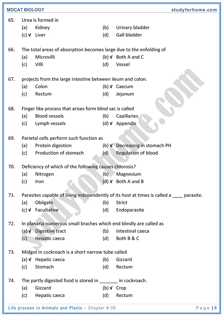
- End result of digestion in stomach is formation of semi-solid mass called chyme.
-
Absorption
- Stomach is involved in the absorption of some chemicals like aspirin and ethanol.
-
Defense/ Immunity
- Mucous membrane and HCl act as barriers against germs.
-
Digestion in Small Intestine
-
- The small intestine consists of three parts i.e. duodenum, jejunum and ileum.
- The entire small intestine is about 6-7m long.
Duodenum
-
- Duodenum is first and the shortest part of small intestine and is about 20-30 cm long.
- When food enters the duodenum, the secretions of pancreas and liver poured into it.
- Duodenum also has its own secretions.
- It acts both as exocrine and endocrine gland.
- Exocrine function of duodenum is secretion of intestinal juice and
- endocrine function is release of secretin and small amount of gastrin hormone.
- Secretin is hormone produced by the action of acidic food on internal mucosa of duodenum. It inhibits production of gastric secretions and promotes production of secretions of liver and pancreas.
- Chyme after neutralization by secretions from liver, pancreas and duodenum is called chyle (liquid).
Role of Liver and Pancreas in Digestion
-
-
Role of Liver:
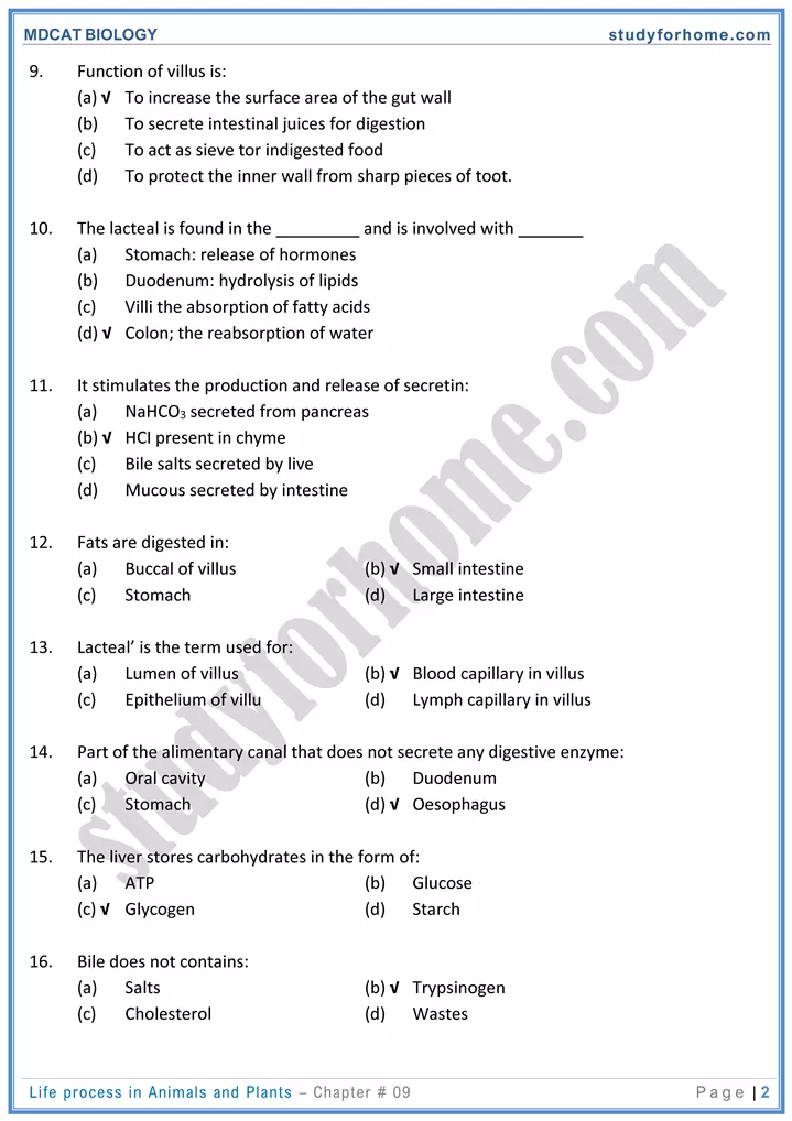
- The liver is the largest internal organ of the body. It has a wide range of functions, including detoxification and production of bile to help digestion.
- Liver secretes bile, which may be temporarily stored in the gall bladder and release into duodenum through bile duct.
-
Composition of Bile
- The bile is green watery fluid consist of bile salts (sodium glycocholate and sodium glycocholate and sodium taurocholate), cholesterol, lecithin, mucus, cellular debris and bile pigments (bilirubin and biliverdin), which are formed from the breakdown of hemoglobin in the liver.
-
Role of Constituents of Bile
- Bile salts reduce surface tension of the fat globules and emulsify them into small droplets and thus increase their total surface area. It is then easily digested by water-soluble lipase.
- Intestinal bacteria convert bilirubin into pigments that give the feces its characteristic brown colour. Some of these pigments are absorbed from the intestine, modified in the kidneys and excreted in the urine, contributed to the characteristic yellowish colour of the urine.
- If bile pigments are prevented from leaving digestive tract, they may accumulate in blood, causing a condition known as jaundice.
- Cholesterol may precipitate in the gall bladder to produce gall stones, which may block the release of bile.
-
Large Intestine
- Large intestine is last pan of alimentary canal. It is divided into caecum, colon and rectum.
- The junction between ileum and large intestine is ileocecal junction gaurded by ileocecal
- sphincter.
-
Caecum:
- Caecum is a blind sac and proximal end of the large intestine. It is the structure the small and large intestines meet.
- Attached to the caecum is a small, blind and finger-like tube which is called appendix It is measured about 9-10cm in length and its wall contains many lymph nodules. inflammation of appendix is called appendicitis.
-
Colon:
- Colon is longest pan of large intestine measuring about.I.5-1.8 m. It is further divided into
- ascending, transverse, descending and sigmoid colon.
- The materials that pass from small intestine to large intestine contain a large amount of water, dissolved salts and undigested material.
-
Rectum
- It is the last part of large intestine where feces are temporarily stored and rejected through anus at intervals.
- Anus is surrounded by two sphincters. The internal anal sphincter is of smooth muscles and outer anal sphincter is of striped muscles.
- Defecation reflex is involved in emptying of rectum from feces. It is generated when rectum is filled with feces. It is consciously controlled in individuals other than infants.
-
Gaseous Exchange/Respiratory system
Anatomy of Human Respiratory System
-
- The human respiratory system can be divided into two regions e.g., upper respiratory
- tract and lower respiratory tract.
- Upper respiratory tract includes nostrils, nasal cavity and pharynx.
- Lower respiratory tract includes the larynx, trachea, bronchi, and lungs.
- Many structures of human respiratory system form the air passage way which provide the path through which air enters or leaves the lungs.
- It consists of following components in sequence:
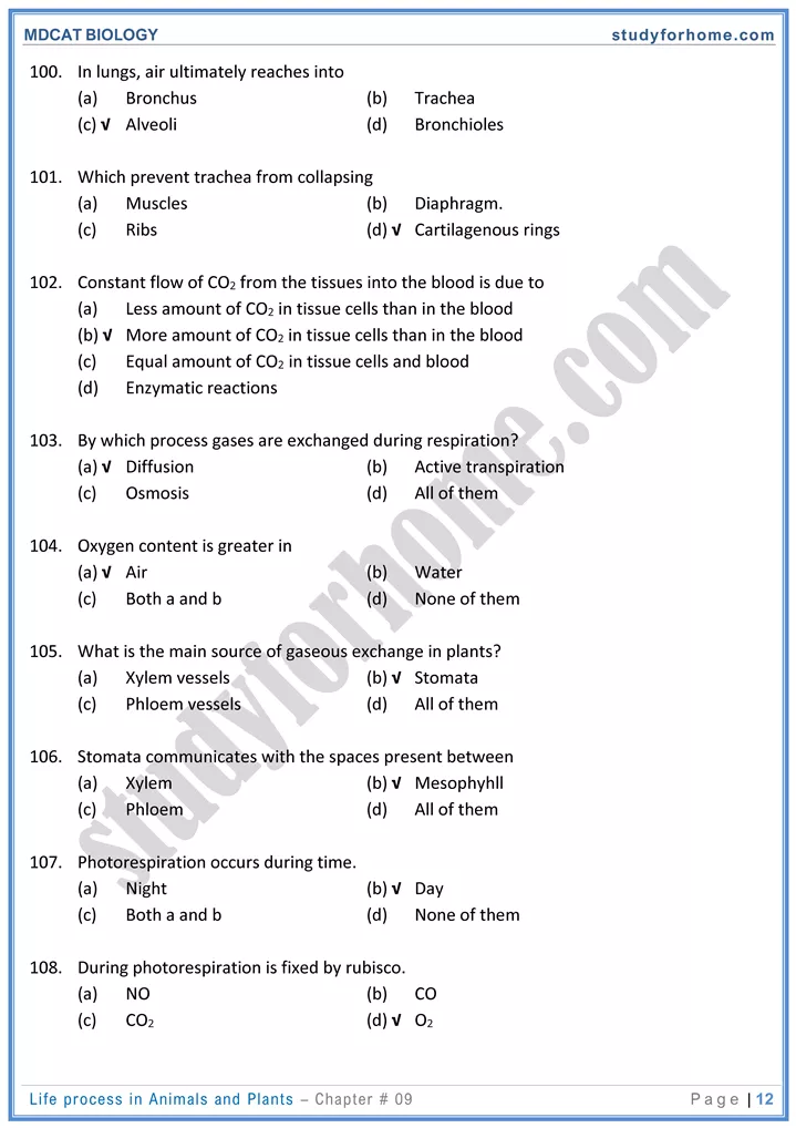
- Nostrils → Nasal Cavities → Pharynx Larynx → Trachea → Bronchi → Terminal
- Bronchioles → Respiratory Bronchioles → Alveolar Ducts → Alveolar Sacs
Mechanism of Breathing
-
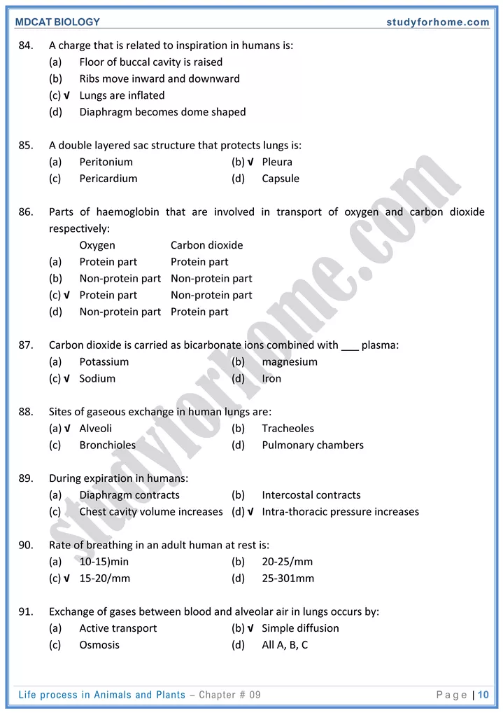
- Breathing is a process by which fresh air containing oxygen is pumped into the lungs and air with more carbon dioxide is pumped out of lungs.
- During rest, breathing occurs rhythmically at the frequency of 15-20 times/min in humans and it can increase to 30 times/min during exercise.
- Lungs are spongy in nature. They themselves neither draw in air nor push it out.
- The floor of the chest cavity is diaphragm which is a sheet of skeletal muscle. It separates the thoracic cavity from abdominal cavity.
- Walls of the chest cavity are composed of ribs and intercostal muscles. There are two sets of intercostal muscles between each pair of ribs: the external intercostal and internal intercostal muscles. The muscle fibres run diagonally but in opposite direction.
Control of Breathing
-
- Normally, breathing is controlled involuntarily but within limits the rate and depth of the breathing are also under voluntary control as shown by the ability to hold the breath.
- Involuntary control of breathing is carried out by a breathing centre located in medulla oblongata of the brain.
- The ventral (lower) portion of the breathing centre acts to increase the rate and depth of inspiration and is called inspiratory centre.
- The dorsal (top) and lateral (side) inhibit inspiration and stimulate expiration. These regions form the expiratory centre.
- The breathing centre communicates with the intercostal muscles by way of the intercostal nerves and with the diaphragm by way of the phrenic nerve. Rhythmic nerve impulses to the diaphragm and intercostal muscles bring about ventilation movements.
- Voluntary control is also used during forced breathing, speech, singing, sneezing and coughing. When such control is being exerted, impulses originating in the cerebral hemisphere ass to the breathing centre which then carries out the appropriate action.
Transport of respiratory gases
-
- Intake of oxygen and release of carbon dioxide by blood passing through capillaries of alveoli is brought about by the following factors:
- Diffusion of oxygen in and carbon dioxide out occurs because of the difference in partial pressures of these gases.
- Within the rich network of capillaries surrounding the alveoli. Blood is distributed in extremely thin layers and therefore, exposed to large alveolar surface.
- Blood in lungs is separated from alveolar air by extremely thin membranes of the capillaries and alveoli.
- Gaseous exchange follows principles of diffusion.
- This exchange occurs due to difference in partial pressure of gases.
Transport of Oxygen
-
- In humans, the transport of oxygen is done by two ways:
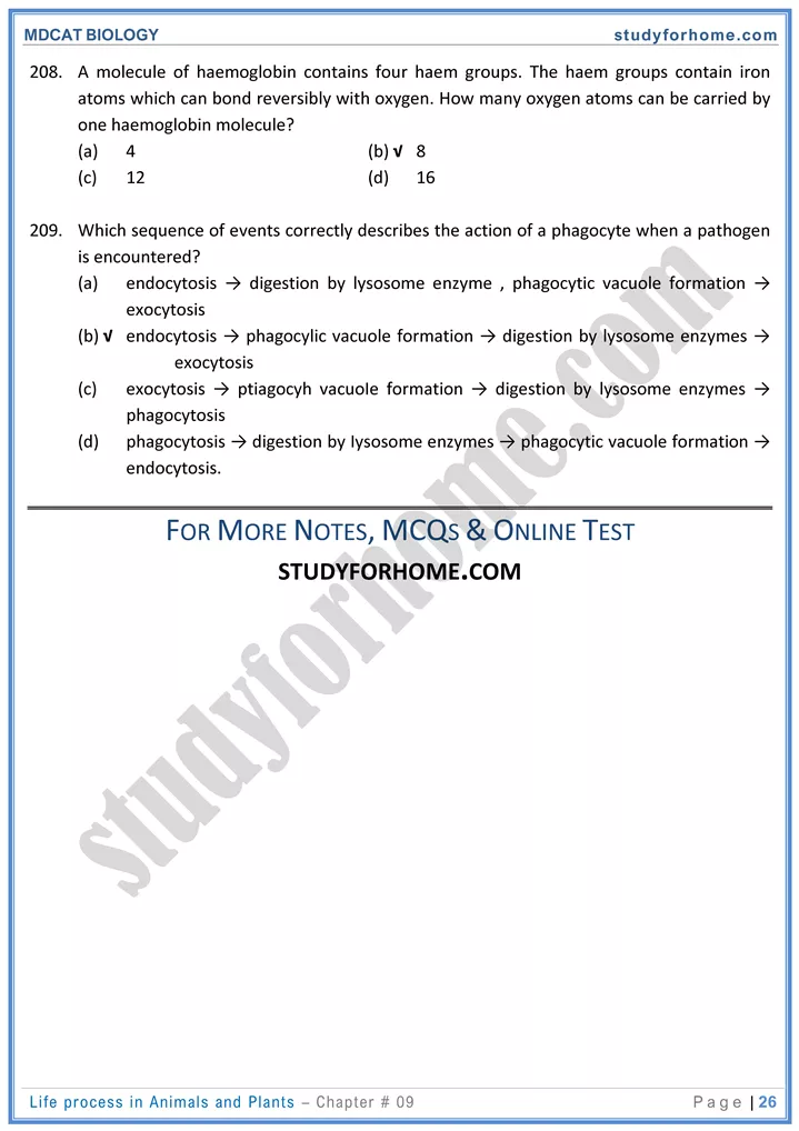
- Approximately, 97% of the oxygen is carried by RBCs as oxyhaemoglobin. Haemoglobin is one of the most important respiratory pigment and acts as an efficient oxygen carrier.
- 3% is transported as dissolved oxygen in the plasma.
Role of Respiratory Pigments
-
- Respiratory pigments are colored molecules, which acts as oxygen carriers by binding reversibly to oxygen. The most important respiratory pigments in humans hemoglobin and myoglobin.
Respiratory volumes
-
- Breathing occurs in cyclic manner due to the movements of the chest wall and the lungs.
- The resulting changes in pressure, causes changes in lung volumes i.e., the amount of air, the lungs are capable of occupying.
- Respiratory volume is the amount of air inhaled, exhaled and stored within the lungs at any given time.
- In an adult human being, when the lungs are fully inflated the total inside capacity of lungs is about 5-6 liters. This is called total lung capacity.
- The amount of air which is inhaled or exhaled at rest is called tidal volume. Its average value is about 500ml.
- The amount of extra air inhaled during a deep breath is called inspiratory reserved volume. This can be as high as 3000ml.
Transport in Plants
Uptake and Transport of Minerals
-
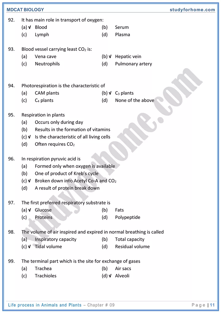
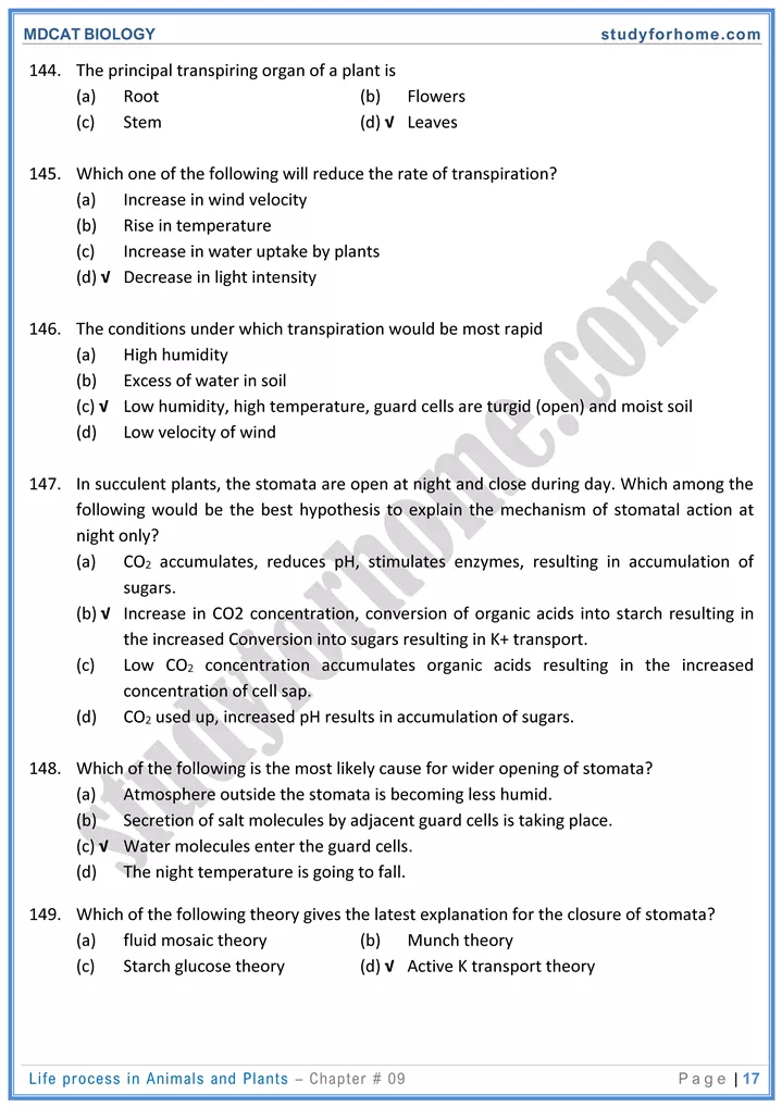
- The roots of a plant not only anchor the plant body in the soil, but also absorb minerals and water from the soil. These nutrients are first absorbed by roots epidermal cells from where these are moved to the xylem and then through xylem, these materials are ultimately reached to the leaves.
- There are three types of nutrients needed by the plants, carbon dioxide, water and minerals besides light to carry out photosynthesis.
- The rate of absorption of individual mineral which is independent of rate of absorption of water molecules is determined by:
- Concentration both inside and outside of the root cells.
- The ease with which it can passively penetrate cell membrane.
- Extent to which carrier molecules and active absorption is involved.
- To get these materials, roots must provide large surface area for absorption, which is achieved by extensive branching. The roots bear a dense cluster of tiny hair-like structures, which are extensions of epidermal cells of roots. These are called root hairs, which are in fact the sites where most of the uptake of water and minerals takes place.
- It has been estimated that out of the total surface area provided by the roots, 67% is provided by the root hair.
- Plants are able to synthesize all their required compounds, with the help of the minerals and H2O from soil, CO2 from air, and light energy.
- Most of the minerals enter the root hairs or epidermal cells of roots along with water in bulk flow, but some are taken in by diffusion, facilitated diffusion, or active transport.
Mineral Absorption by Roots
-
- The minerals available to plants for absorption are dissolved in the soil water. Their concentration varies according to the fertility and the acidity of the soil, besides other factors.
- When the soil minerals are not in solution but are bound by ionic bonds to soil particles, they are not available to plants.
Processes Involved in Absorption by Roots
-
- The uptake of minerals by root cells is a combination of passive uptake and active uptake, involving the use of energy in the form of ATP.
Water Potential
-
- Water molecules possess kinetic energy which means that in liquid or gaseous form, they move about rapidly and randomly from one place to another. So, greater the concentration of the water molecules in a system, the greater is the total kinetic energy of water molecules. This is called water potential and is represented by (Yw).
- (Yw) of a medium is directly proportional to the concentration of water in that medium; therefore, pure water has highest value of (Yw) which is designated a value of zero.
- (Yw) of all the solutions or the cells must be less than zero i.e., in negative range.
Uptake of Water by Roots
-
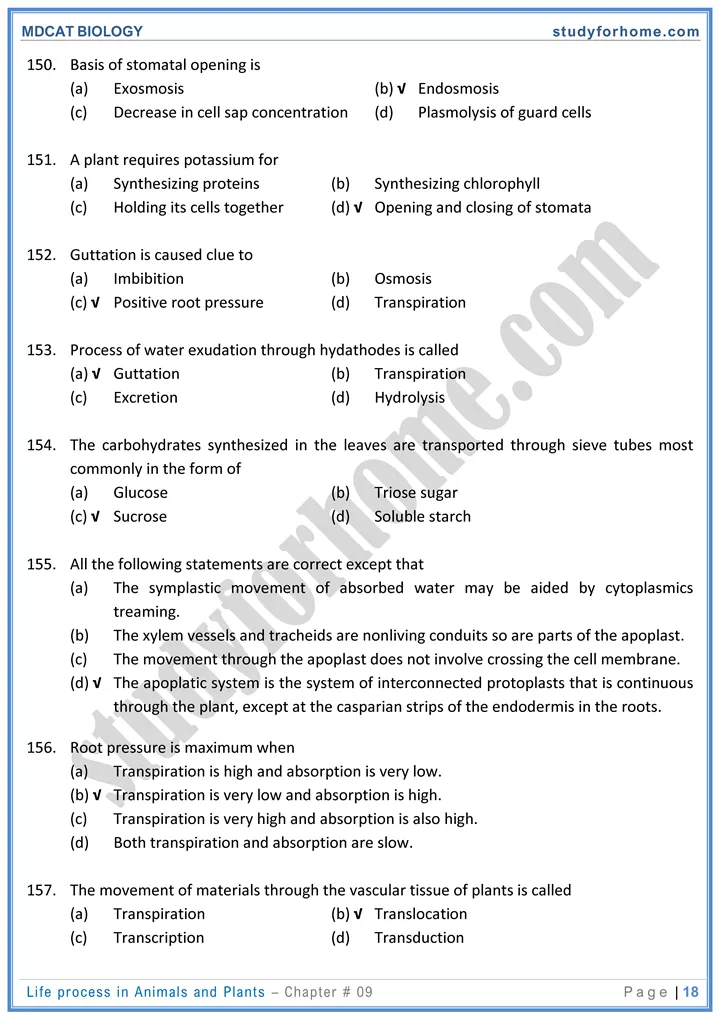
- The cell wall of epidermal cells of roots is freely permeable to water and other minerals, while cell membrane is differentially permeable. From root hairs, water molecules enter the epidermal cells by osmosis i.e., along the concentration gradient.
- Is passes through cortex, endodermis, Pericycle, and reach the xylem vessel.
- The movement of water into the cells in called endosmosis while the movement of water out of the cell is called exosmosis.
- There are three pathways taken by water to reach the xylem tissues i.e. apoplast, symplast and vacuolar pathway.
Apoplast Pathway
-
- This involves system of adjacent cell walls, which is continuous throughout the plant roots.
- In the roots, apoplast pathway becomes discontinuous in the endodermis due to the presence of casparian strips which is a band of suberin and lignin, bordering four sides of root endodermal cells.
Symplast Pathway
-
- It is an important pathway of water movement in which movement of cell sap involves the cytoplasmic connection of adjacent cells.
- The cytoplasm of neighboring cells (protoplast) is linked with one another by plasmodesmata.
Vacuolar Pathway
-
- In this pathway, water moves from vacuole to vacuole through neighboring cells, crossing the symplast and apoplast and moving through membranes and tonoplast by osmosis. It moves down a water potential gradient.
Ascent of Sap
-
- Water and minerals are absorbed by the roots epidermal cells from where these substances are first move to the root xylem and then to the leaves. This upward movement of water and dissolved minerals towards the leaves through the xylem tissue is called ascent of sap.
Translocation of Organic Solutes
-
- The cells of phloem that transport sugars and organic material throughout the plant are called sieve elements. These cells are associated with one or more companion cells.
- The movement of prepared organic solutes to different parts of the plant body through phloem tissue is called translocation of organic solutes.
- Sieve tube and companion cells are in communication with each other by plasmodesmata.
- Companion cells supply ATP and proteins to the sieve tube.
Transport in Man/Cardiovascular system
Blood
-
- The weight of blood in our body is about 1/12″ of our body.
- Blood is the form of fluid connective tissue in which dissolved nutrients, respiratory gases, hormones, and wastes are transported through the body.
- The normal pH of blood is 7.4.
- made up of two main components i.e. plasma and cells or cell like bodies.
Structure of Human Heart
-
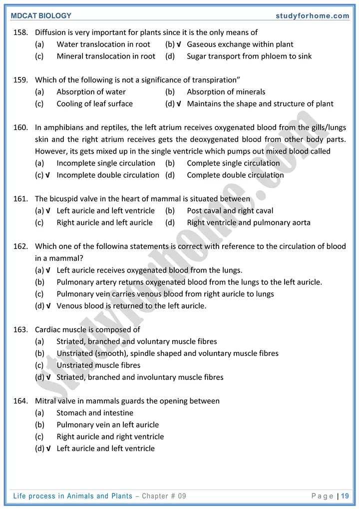
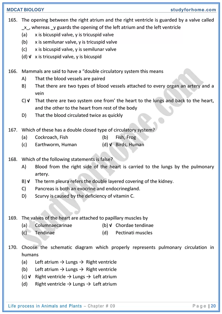
- Human heart is hollow, fibromuscular organ. Adult heart has shape of cone.
- The human heart is located in the chest cavity between lungs slightly left of the sternum.
- The heart contracts automatically with rhythmicity, under the control of the autonomic nervous system.
- Base of heart extends to second intercostal space and apex of heart is in fifth intercostal space, approximately 9 cm to left of midline.
Heart Walls:
-
- Epicardium is a thin serous membrane comprising of smooth outer surface of heart
- The wall of the heart is composed of three layers: Epicardium, Myocardium and Endocardium.
- Myocardium of heart is made up of special type of muscles, the cardiac muscles. Their arrangement and mechanism of contraction is essentially same as skeletal muscles except that they are branched cells. Successive cells are separated by junctions called intercalated discs.
- Endocardium consists of simple squamous epithelium over a layer of connective tissue.
- Heart valves are formed by fold of endocardium.
The Cardiac Cycle
-
- Heart beat involves three distinct stages i.e. atrial systole, ventricular systole and diastole.
- It is the sequence of events which take place during the completion of one heartbeat.
- Relaxed period of heart chambers is called diastole and contraction is called systole.
- One complete heart beat consists of one systole and one diastole and lasts for about 0.8 seconds
- In one’s life, heart contracts about 2.5 billion times, without stopping.
Mechanism of Heart Excitation and Contraction
-
- Specialized strands of interconnecting cardiac muscle tissue that coordinate cardiac contraction constitute the conduction system. The conduction system constitutes the cardiac cycle. Cardiac muscle has an intrinsic rhythmicity.
- The heart will go in beating after it has been cut right out of the body. Cardiac muscles are myogenic i.e., its rhythmic contraction arise from within the muscle itself.
- Heartbeat starts when the sino-atrial node (pacemaker) sends out electrical impulses to the atrial muscles, thus causing both atria to contract. It is located at the upper end of right atrium.
Blood Pressure and Rate of Blood Flow
-
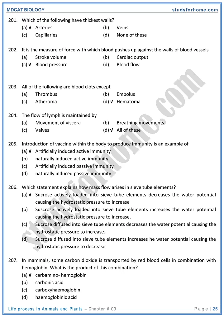
- It is the measure of force with which blood pushes up per unit area against the walls of blood vessels.
- It is measured in mmHg.
- Blood pressure is detected by mechanoreceptors called baroreceptors.
- It is the force that keeps blood flowing from the heart to all the capillary networks in the body.
- The blood pressure is generated by the contraction of ventricles. This is called systolic pressure.
- When the ventricles relax, the atrial pressure is lowest and is called diastolic pressure.
- Blood pressure consistently decreases in the following pathway: Aorta → Arteries → Capillaries → Veins → Vena cava
- The normal systolic blood pressure is 120 mm Hg which is during ventricular systole.
- The normal diastolic blood pressure is 75-85 mm Hg which is during diastole of the heart.
Lymphatic System
- This system is responsible for the transport and returning of material from the tissues of the body to the blood.
- It comprises of lymph capillaries, lymph vessels, lymphoid masses, lymph nodes, lymph and lymphocytes.
- Lymph is the fluid which flows in the system. The fluid in the interstitium is derived by filtration and diffusion from the capillaries. The intercellular spaces in the walls of lymph vessels are larger than those of the capillaries of blood vascular system.
- This fluid is mainly entrapped in the minute space among the proteoglycan filaments. This combination of proteoglycan filaments and the fluid entrapped within them the characteristics of gel and therefore is called tissue gel.
- Approximately 30 liters of fluid pass from the blood capillaries into the interstitial space each day. Whereas only 27 liters pass from the interstitial space back into blood capillaries. The remaining 3 liters of fluid enters the lymphatic capillaries, where the fluid is called lymph, and passes through the lymphatic vessels back to the blood.
- After a fatty meal, the fat globules may make up 1% of the lymph.
- The lymph vessels empty in veins; so lymph is a fluid in transit between interstitial fluid and the blood.
- In addition to water lymph contains solutes such as ions, nutrients, gases and some proteins, hormones, enzymes and waste products.
Immune System
Immunity
-
- The capacity to recognize the intrusion of any material foreign to the body and to mobilize cells and cell products to help remove the particular sort of foreign material with greater speed and effectiveness is called immunity
- The body’s response to foreign particles, such as the production of antibodies directed against a specific antigen, is called an immune response.
Defense Lines
-
- The human body has three lines of defence against microbial attack.
- First and second defense lines are non-specific while 3rd defense line is specific.
- Skin, mucous membrane and blood clot are physical barriers.
- HCI and lysozyme are examples of chemical barriers.
- Phagocytes and lymphocytes are example of cellular/ biological barriers.
