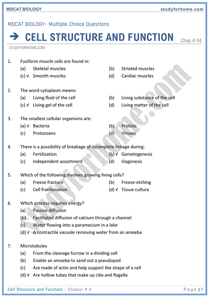
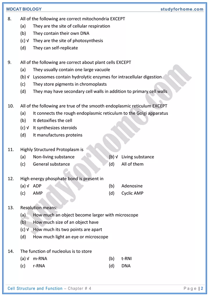
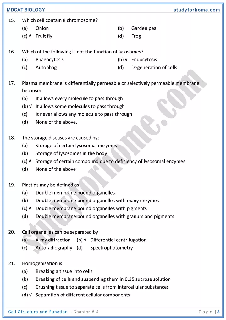
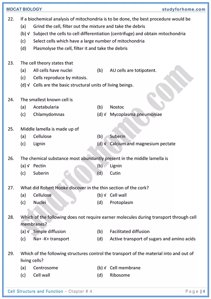
MDCAT Biology – Chap#4 Cell Structure And Function
Microscopes:
Depending upon source of illumination following are the broad types of microscopes.
| Source of illumination | Lenses | Resolution | Type of Substance Studied | |
|---|---|---|---|---|
| Light of Microscope | Visible light | Glass lens | Low | Living |
| X-ray Microscope | X-Rays | Electromagnetic | High | Non-Living |
| Electron Microscope | Beam of electrons | Electromagnetic | High | Non-living |
Terms:
- Magnification: Enlarging the apparent size of an object compared to its actual size
- Resolution: Ability to show two adjacent objects as separate. It refers to clarity and detail of an image.
- Contrast: to distinguish one part of a cell from another. Regarding light microscope contrast is obtained by fixing and staining the material.
Cell:
Basic components of a cell are:
- Plasma membrane
- Cytoplasm
- Nucleus
1. Plasma membrane:
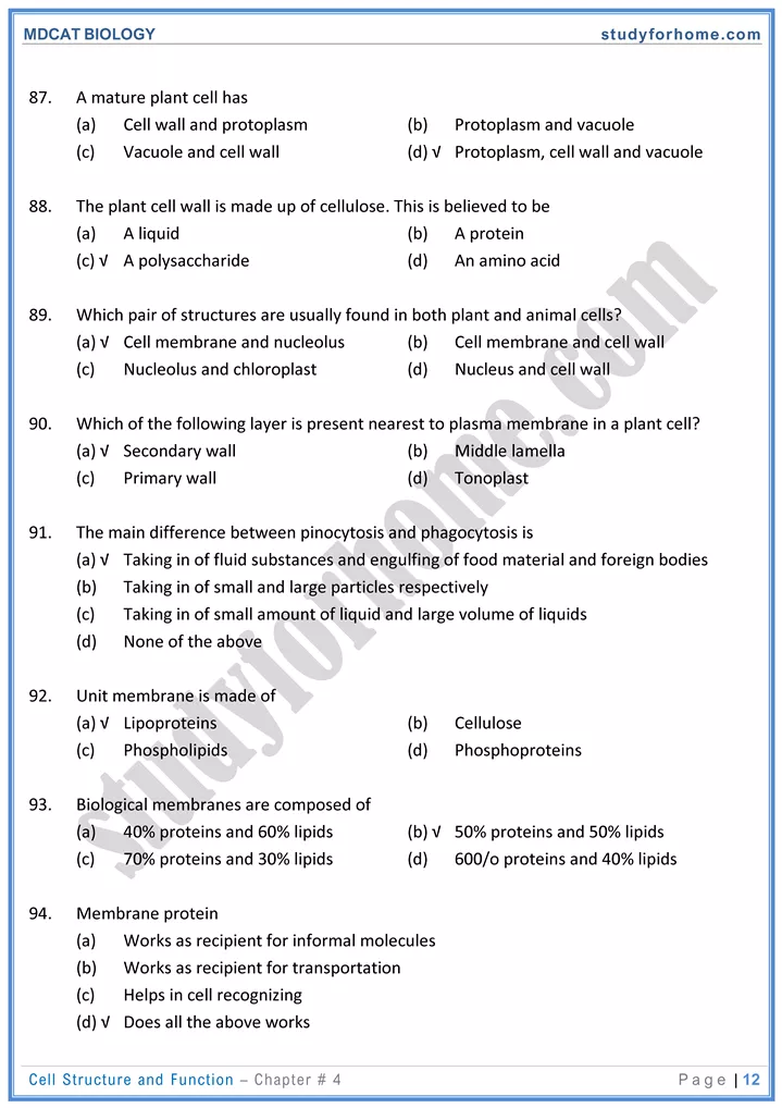
- Composition: 60-80% proteins, 20-40% lipids, some carbohydrates
- Structural organization
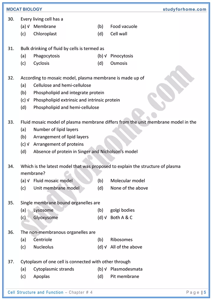
- Unit membrane: This proposed that the lipid bilayer is sandwiched between inner and outer layers of proteins. This basic structure is called unit membrane.
- Fluid Mosaic Model: Recent model shows that proteins are scattered in the lipid bilayer which is in fluid state. Thus fluid refers to lipid bilayer and scattered protein refers to Mosaic in this model
- Peripheral / Extrinsic proteins: these proteins are located on either side of the lipid bilayer.
- Integral / Intrinsic proteins: these proteins are partially I completely imbedded in the lipid bilayer.
- Cell Wall: The outer most boundary of plant cells and some other organisms: It is secreted by protoplasm. It can be differentiated into 3 layers.
- Middle lamella: present as a cementing layer between two adjacent cells. It is actually the first formed cell plate composed of calcium and Magnesium pectate.
- Primary wall: it is composed of cellulose hemicellulose and pectates. It is the first product of protoplasm deposited on either side of Middle lamella.
- Secondary wall: It is formed on inner side of primary wall. In addition to cellulose lignin or other substances may also be present.
- Plasmodesmata: These are minute channels in the cell wall where deposition of cell wall material does not take place so that cytoplasm of adjacent cells remains in contact.
- Being hydrophilic in nature, cell wall can imbibe large amounts of water.
2. Nucleus
A eukaryotic nucleus has following components.
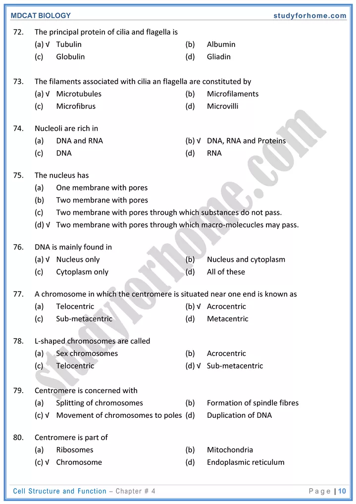
- Nuclear membrane: It is a double membrane surrounding the nucleoplasm /karyo lymph and it has pores in it called nucleopores. It is also called as nuclear envelope.
- Nucleolus: It consists of RNA, DNA and proteins. It is involved in synthesis of ribosomes and RNA. It disappears during cell division and reappears in the daughter cells
- Chromatin network / nuclear reticulum: It is noncompact form of chromosomes.
- Chromatin fibers are composed of DNA associated with proteins
3. Cytoplasm
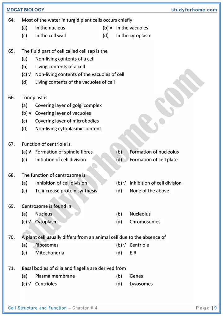
- The fluid between plasma membrane and nuclear membrane is cytoplasm. Its ground substance is cytoplasm.
- Glycolysis takes place in cytoplasm.
Cytoplasmic Organelles:
Organelles refers to division of labor in a cell. Eukaryotes and prokaryotes differ in following aspects.
| Prokaryotic Cell | Eukaryotic Cell |
|---|---|
| Mitochondria, endoplasmic reticulum, plastids, golgi bodies can never be present in a prokaryotic cell. Genetic material is diffused or not surrounded by membrane. Nucleoid is the region where nuclear material is present. Ribosome is of 70s. Cell wall is made up of peptlidoglycan / murein. Molecular complex of cell wall is regarded as sacculus e.g. Bacteria and blue green algae. | All double membrane bound organelles are present depending upon the type of cell i.e. plastids in plant cells only. Genetic material is surrounded by double membrane called as nuclear membrane or envelope. 80s ribosomes are present. In plant cells, cell wall is made up of cellulose e.g. protests, fungi plants and animals. |
Endoplasmic Reticulum:
It is a network of tubules extending from nuclear membrane to plasma membrane. They serve functions of support, secretion, transport, synthesis and storage in a cell. Depending upon presence / Absence of ribosomes on it. There are two types of endoplasmic reticulum.
Rough / granular endoplasmic reticulum: Ribosomes are attached to it. REP are associated with protein synthesis.
Smooth / granulate endoplasmic reticulum: Its surface is free and is associated with synthesis of lipids like sex hormones, etc. It also stores ions in ii responsible for muscle contraction.
ER also provides structural support to the cell.
Ribosomes:
- Ribosomes are non-membrane bound organelles and it has two subunits. The larger subunit sediments at 60s and smaller subunit at 40s. Two sub-units attach to form 80g unit.
- During protein synthesis ribosomes are attached to mRNA forming a polysome.
- They are responsible for granular appearance of cytoplasm.
- They are synthesized in nucleolus and they are protein factories of a cell.
SINGLE membrane bound organelles:
| Lysosomes | Peroxisome | Glyoxisomes |
|---|---|---|
| Present in plant and animal cells. Hydrolytic enzymes are present. Responsible for intracellular digestion autophagy etc. Also called a suicide sacs | In plant and animal cell. Oxidative enzymes are present. Responsible for detoxification of substances. | Present only in plants Enzymes converting fatly acids to carbohydrates are present. Present in lipid rich germinating seeds e.g. castor beans. soya beans etc. |
Formation of Ivsosomes and its various forms:
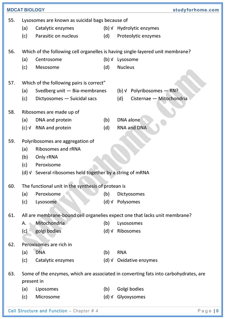
- Hydrolytic enzymes from RER or ribosomes are packaged by goIgi bodies in the form of vesicle. This vesicle is called primary lysosomes.
- When cell Debrisor any substance is engulfed by lvsosome, this heterogeneous structure is called secondary lysosome.
- after digestion, remaining component is called residual body
Cytoskeleton:
In addition to endoplasmic reticulum following structures are responsible for skeletal support.
| Microtubules | Intermediate filaments | Microfilaments |
|---|---|---|
| Hollow tubules having 25nm diameter. | Solid strands having 8 to 1mm diameter. | Solid strands having 7nm diameter. – |
| Present in cilia, flagella, spindle fibres and cenrioles | Maintains shape of cell and keeps organelles in its position. | Responsible for internal cell motion. |
| Made up of protein tubulin. | – e.g. muscle contraction. cyclosis and formation of pseudoopodia. |
Mitochondria / oxysomes :-
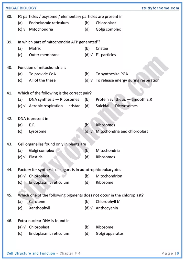
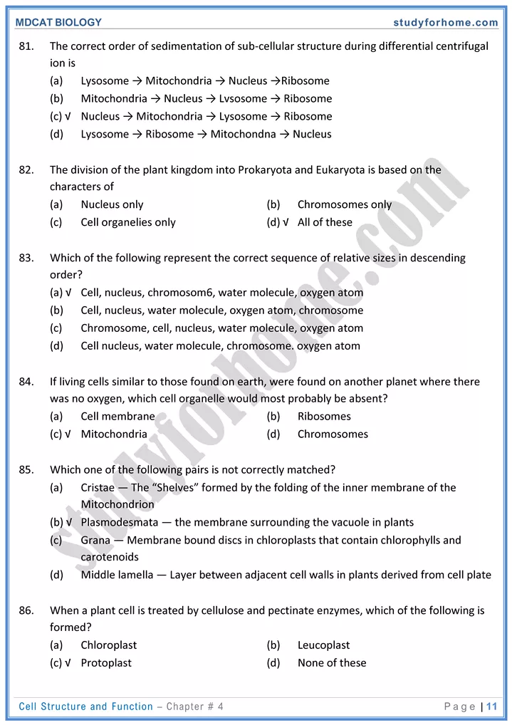
They are double membrane bounded organelles present in all eukaryotic cells. The outer membrane is smooth whereas inner membrane has folds having knob like structures called as F1 particles matrix is the fluid present in it.
- F1 particles are associated with ATP synthesis.
- It has its own circular DNA and ribosomes which are of primitive type
- The are called as power houses of a cell
- Various metabolic processes like Krebs cycle, aerobic respiration, fatty acid oxidation occur in it.
Plastids :-
- Present only in plant cells. There are three types of plastids, all are derived from pro plastid.
Chloroplast:
- It contains green pigments. fluid present in it is called stroma. Stroma has enzymes for dark reaction.
- A single stack of membrane containing pigment is thylakoid, stacks of thylakoid refers to granum.
- Grana are attached to each other via non green part called intergranum.
- Light energy is trapped in the membrane of grana and ATP formation occurs. B) Chromoplast:- plastids other than green are chromoplast
- Leucoplast: plastids having no color are called leucoplast they are present in underground parts of plant.
- Golgi bodies (dictyosomes.)
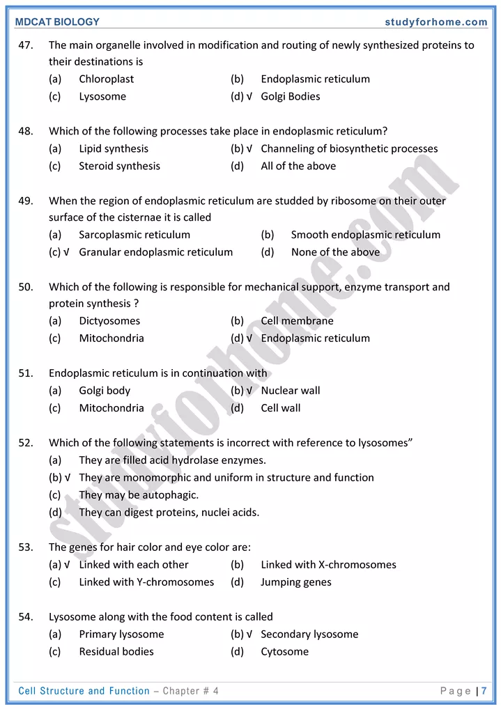
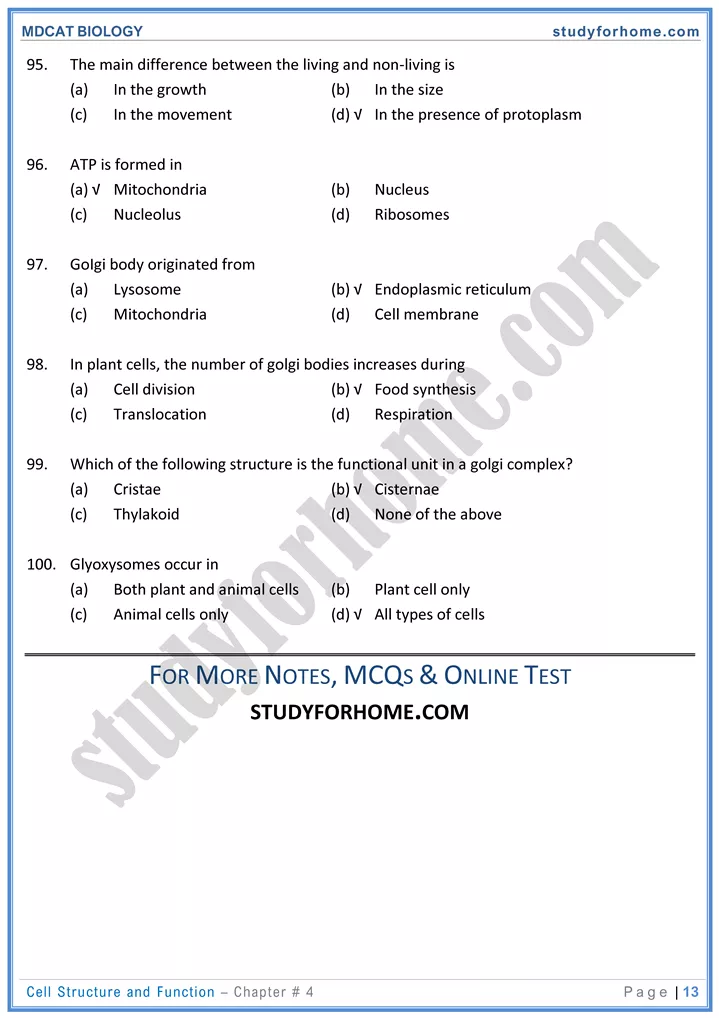
- They are formed from budding off the smooth endoplasmic reticulum. They are flatrened stacks of membrane bound;- Sacs called cisternae.
- Stack along with vesicles comprise golgi apparatus
- They are responsible for packaging, glycosylation of proteinslipids and cellular secretions.
- cell plate (later middle Iamella in plant cell is formed by golgi bodies
Endomembrane System:-
- In a eukaryotic cell, endoplasmic reticulum golgi bodes / vesicles, comprise endomembrane system.
- Synthesized product from ER can directly secreted from it or it could send to golgi bodies for further packaging prior to secretion.
Same facts
- Spherical tubular membrane I flattened stacks of ERI Golgi bodies both are called as cisternae
- Cristae are folds of inner mitochondrial membrane
- Depending upon the position of centromere chromosomes are divided into Telo. Acreo submeta and metacentric.
- Factory of ribosome is nucleolus.
- Factory of protein is ribosome.
- Polysome or polyribosome is a complex of am-RNA and ribosomes during protein synthesis.
- Chloroplasts and mitochondria has its own DNA and protein factory. Thus they are self replicating.
