Support & movement – Chap 12 – Biology MDCAT
-
In Animals:
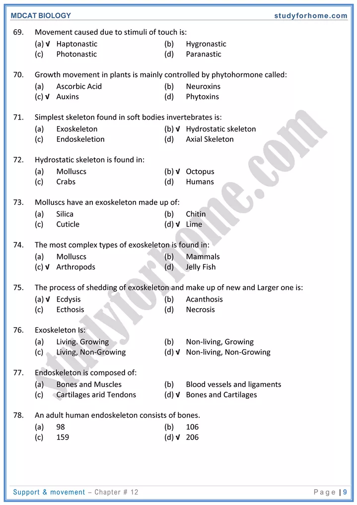
- Hydrostatic skeleton consists of fluid held under pressure. E.g. in hydra, jelly fish, sea anemone, octopus etc.
- This helps jet propulsion in hydra and octopus.
- This skeleton cannot serve function of protection.
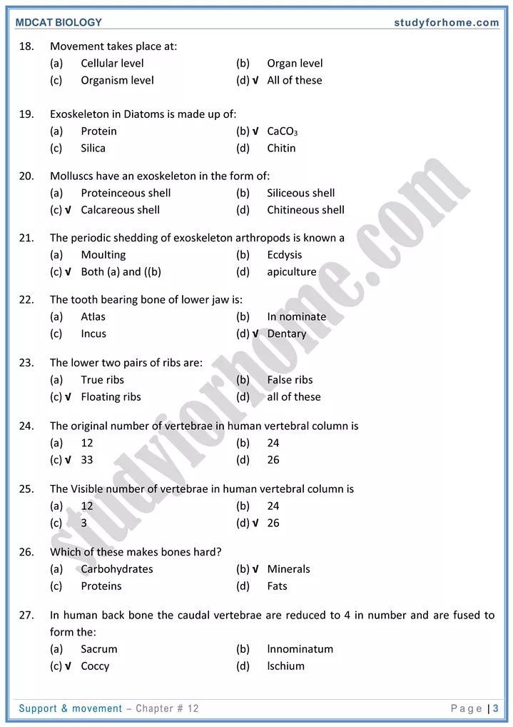
- Exoskeleton is hard non-living outer covering of body secreted by epidermal cells. It Is silicaceous in diatoms, made up of CaCO3 I lime in molluscs and chitinaceous in arthropods.
- Gradual shedding of exoskeleton and replacing it with new one is called ecdysis / Moulting.
- Endoskeleton in vertebrates is made up of bones / cartilage which is living connective tissue.
- Spicules of sponges and calcareous plates of echinoderms are dead structures.
-
Human Skeleton:
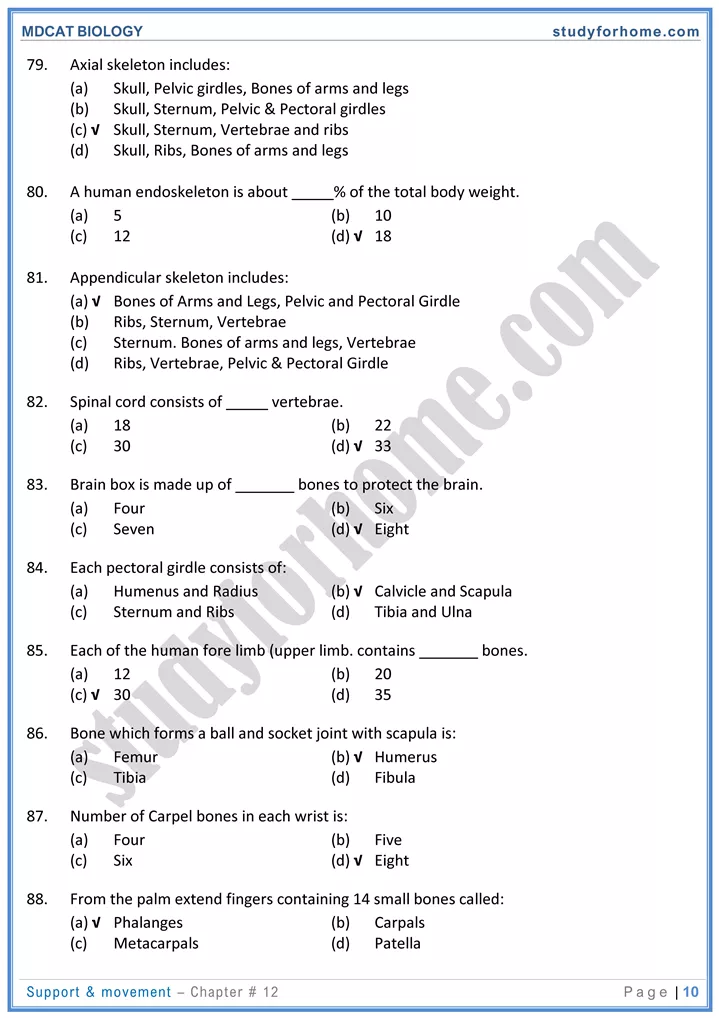
- Axial skeleton includes skull, vertebral column, sternum and ribs.
- Appendicular skeleton includes girdles and limbs. They work as levers.
-
Bones
- Axial Skeleton
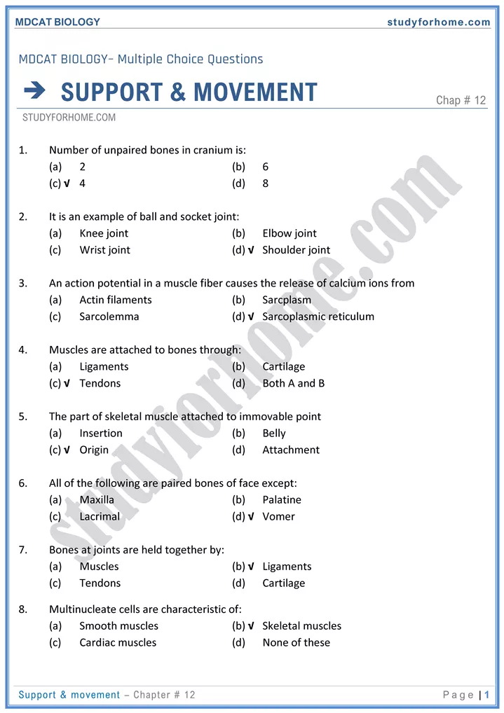
- Cranium (brain box) = 8 bones, facial bones 14 bones. Lower jaw bone is called Dentary.
- Original number of vertebrae →, 33 after fusion 26.
- Cervical vertebrae →, 7, thoracic vertebra 12, lumbar vertebra = 5, sacral →5, caudal →
- Ribs → 12 pairs (last two pairs are called floating ribs.
-
Appendicular Skeleton
- Pectoral girdie → scapula and clavicle. Scapula has glenoid cavity.
- Clavicle / collar bone attaches appendicular skeleton → axial skeleton.
- Upper limb →, 30 bones; human, radius, ulna, carpal = 8 bones (forming wrist),
- metacarpals (palm bones) = 5, phalanges (fingers and thumb) = 14 in number.
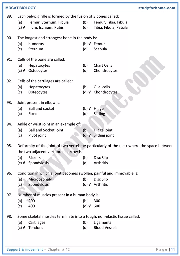
- Pelvic girdle formed of innominate bone (ilium. ischiurn, pubis) forms cavity acetabulum.
- Leg bones, femur, tibia, fibula, tarsals (7), metatarsals (5), phalanges (14).
-
Joints
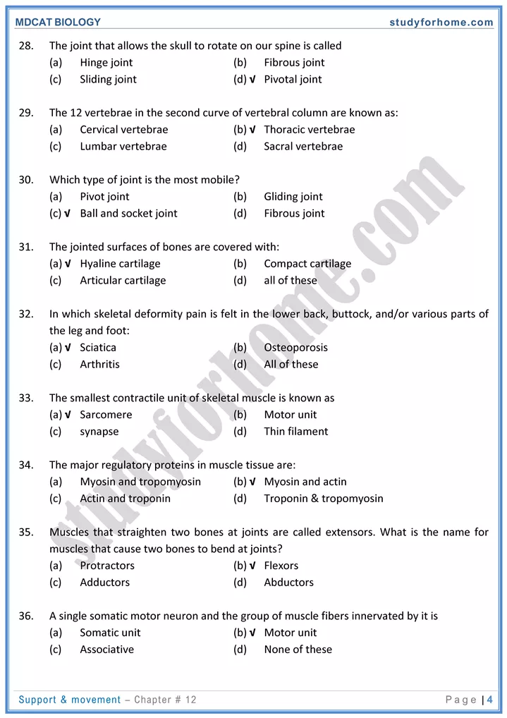
- Hinge jointg. elbow, knee, finger joints etc.
- Ball and socket jointg. shoulder and hip joints.
- Pivot joints between skull and vertebral column.
- Sliding jointsg. joints between bones of wrist and ankle.
- Gliding jointsg. joints between vertebrae.
- Partially moveable jointsg. joints between ribs and vertebral column.
- immovable joints/suturesg. joints between bones of skull and vertebral column.
- Fibrous jointsg. in skull bones.
- Cartilaginous jointsg. joints between vertebrae.
- Synovial jointsg. shoulder & hip
-
Divisions of Human Skeleton:
- There are about 350 bones in an infant and 206 in adult human. The human skeleton is primarily divided into two divisions i.e., axial skeleton (80 bones) and appendicular skeleton (126 bones).
- Appendicular skeleton is associated with extremities and consists of pectoral girdle with forelimbs and pelvic girdle with hind limbs.
-
Axial Skeleton:
- Axial skeleton provides basic framework of body and includes those skeletal parts which are present along the central axis of the body, like skull, vertebral column, rib cage and sternum.
- Primary function of skull is protection of brain, while vertebral column provides protection to spinal cord. It has four curvatures.
-
Types of Joints:
- A joint or articulation is a place where two or more bones and cartilage come together.’ The scientific study of the structure and function of joints is called arthrology.
- Joints have two fundamental functions which are:
- They give mobility to the skeleton.
- Hold the skeletal parts together.
-
Classification of Joints:
- The joints mainly are classified as on the basis of structure and function. Structural classification of joints is based on the material binding the bones together and whether or not a joint cavity is present while functional classified is based on the amount of movement allowed by them.
- structurally, there are three types of joints; fibrous (immovable), cartilaginous (slightly movable), and synovial joints freely movable.
- It is the inflammation of joints characterized by the degeneration and damage to the joint tissue.
- The typical symptoms of arthritis include pain after walking which may later occur even at rest, creaking sounds in joints, difficulty in getting up from a chair and pain on walking up or down stairs.
- Osteoarthritis is a progressive disease in which the articular cartilages gradually soften and disintegrate. It affects knee, hip and intervertebral joints.
- Rheumatoid arthritis is the result of an auto-immune disorder in which synovial membrane becomes inflamed due to faulty immune system.
- Gouty arthritis results form a metabolic disorder in which an abnormal amount of uric acid is retained in the blood and sodium urate crystals are deposited in the joints.
-
Types of Muscles:
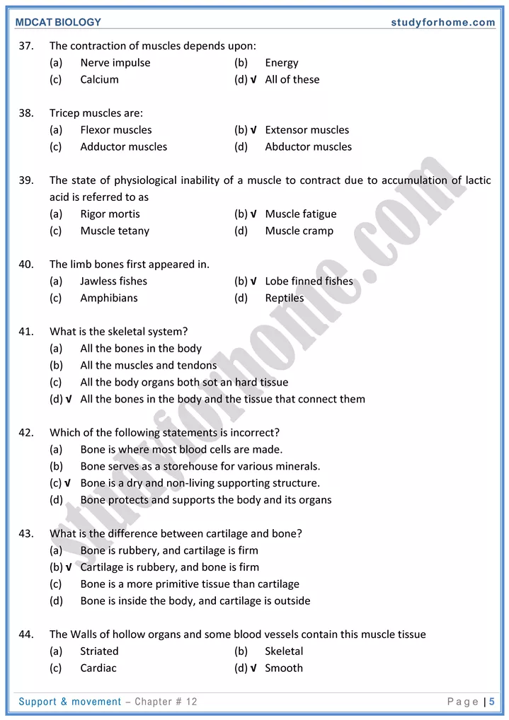
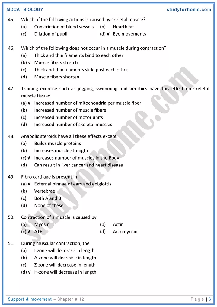
- Muscles are the specialized tissues of mesodermal origin that can undergo contraction and relaxation and provide movements to body parts or the whole body. The study of the muscles is called myology.
- The most distinguishing functional characteristic of muscles is their ability to transform ATP into mechanical energy. In doing so, they become capable of exerting force.
- Earliest forms of muscles to be evolved are smooth muscles which are present throughout animal kingdom. Cardiac muscles and skeletal muscles are found only in vertebrates.
- Cardiac muscles have intercalated disc.
- They also function to hold body parts in postural positions, movement of body fluids and heart production.
-
Structure and Ultra-Structure of Skeletal Muscle:
- The muscles that are attached to the skeleton and are associated with the movement of bones are called skeletal muscles.
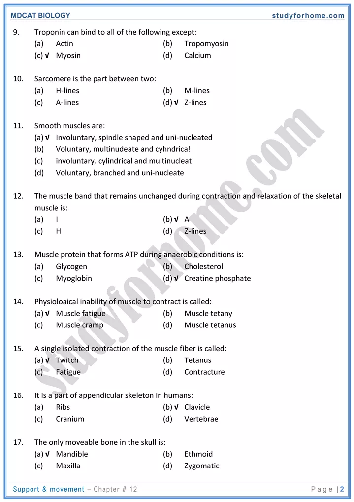
- The skeletal muscles are consciously controlled and therefore, are called voluntary muscles.
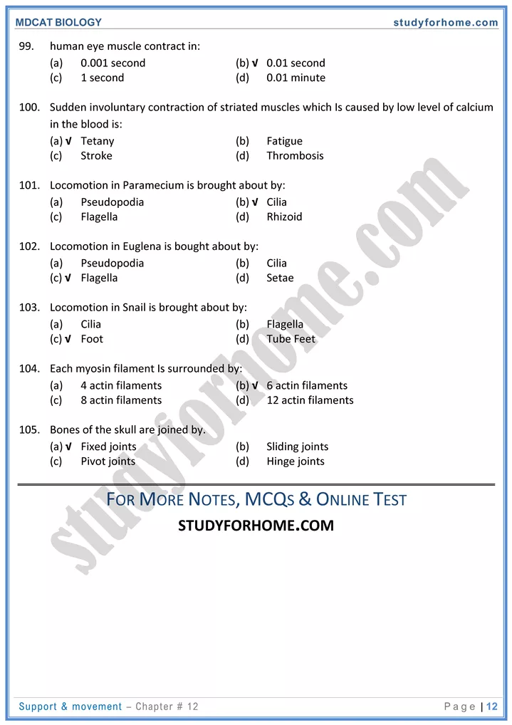
- Generally, each end of the entire muscle is attached to bone by a bundle of collagen, non-elastic fibers known as tendons.
-
Muscle Bundle:
- Muscles bundles ate also called as muscle fasciculi.
- These are bounded by a connective tissue called perimysium.
- Muscle bundles are further composed of muscle fibers or cells.
-
Muscle Fibers:
- Each muscle fiber is a long, cylindrical and huge cell with the diameter of about 10-100 μm and contain multiple oval nuclei which are arranged just beneath its sarcolemma.
- The sarcolemma of muscle fibres penetrates deep into the cell to form a hollow elongated tube, the transverse tubule / T-tubule.
- Sarcoplasm of the muscle fiber is similar to the cytoplasm of Other cells, hut it contains usually large amount of stored glycogen and unique oxygen binding protein e.g. myoglobin.
- sarcoplasmic reticulum is continuous system of sarco-tubules extending throughout the sarcoplasm around each myofibril. It is like endoplasmic but devoid of ribosomes.
- These act as store-house of Ca+2.
- Each muscle fiber further contains large number of myofibrils.
-
Myofibrils:
- When viewed in high magnification, each muscle fibre is seen to contain a large number of myofibrils, 1-2 pm in diameter that run in parallel fashion and extend entire length of cell. Bundles of these fibrils are enclosed by the sarcolemma.
-
Banding Pattern:
- Myofibril has series of dark and light bands.
- Each dark band is called A-band because it is anisotropic i.e., it can polarize visible light.
- The light band is called I-band because it cannot polarize visible light. It gives cell as whole its striped appearance.
- Each A-band has a lighter stripe in its mid-section called H-zone. The H-zone is bisected by a dark line called M-line. The I-band has mid line called Z-line.
- A sarcomere is the region of a myofibril between two successive Z-lines and is the smallest contractile functional unit of muscle fibres.
-
T-Tubules, T-System & Triad:
- The sarcolemma of muscle fiber Cell penetrates deep into the cell to form hollow elongated tube, the transverse tubule or T-tubule, the lumen of which is continuous with the extracellular fluid.
- Thousands of T-tubules of each muscle cell are collectively called as T-system. It extends and encircles the myofibril at the level of Z-line or A-I junction.
- The T-tubule and the terminal portion of the adjacent envelope of sarcoplasmic reticulum form triads at regular interval along the length of the fibril.
-
Effect of Exercise on Muscles:
- The amount of work a muscle does is reflected in changes in the muscle itself. For example;
-
- Increase in size of the muscle
- Increase in its strength
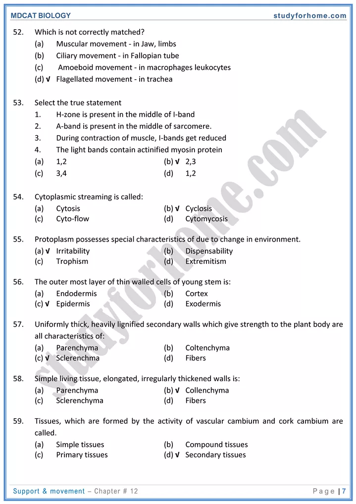
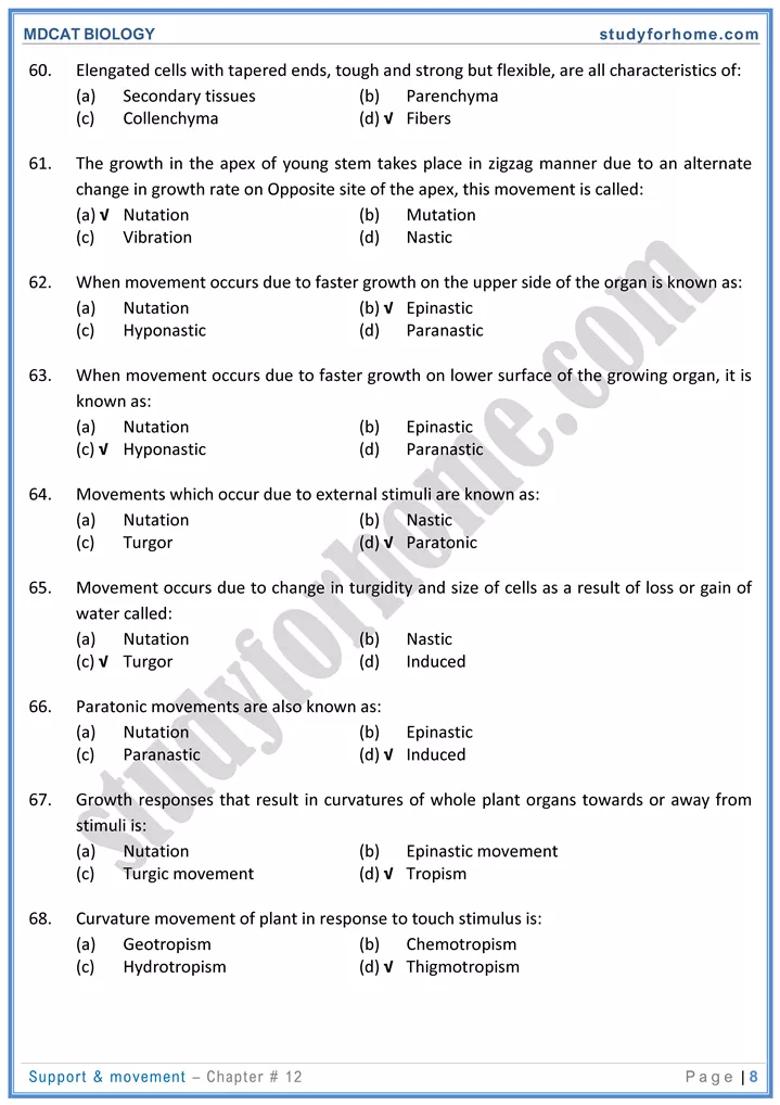
- More efficient and fatigue resistant
- Capillaries surrounding muscle fibers and mitochondria in it increases
- Synthesize more myoglobin
-
- Complete immobilization of muscle leads to muscle weakness and severe atrophy.
